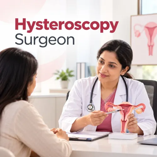


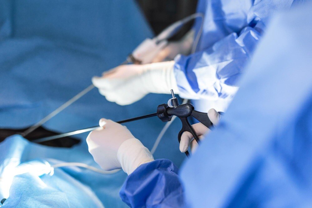
The phrase “hysteroscopy surgery” can describe a camera examination, removal of tissue, or a planned treatment completed during the same session. You will learn what happens from the first consultation through recovery, which questions to ask about anaesthesia and facility arrangements, and how pathology results guide the next step.
Key takeaways
- Bring your bleeding, pregnancy, surgery, medicine and allergy history to consultation.
- Hysteroscopy can identify and treat polyps, fibroids, adhesions and retained tissue.
- Ask whether your procedure is diagnostic or operative and what anaesthesia it requires.
- Confirm how tissue samples will be labelled, tested and when results will be shared.
What happens before hysteroscopy is scheduled?
Scheduling starts with a consultation that determines whether hysteroscopy is appropriate and whether the planned procedure is diagnostic or operative. Discuss your menstrual pattern, bleeding duration, pain, pregnancy and infertility history, previous miscarriages, Caesarean or uterine procedures, medicines, allergies, anaesthesia reactions and medical conditions.
The clinician may perform a pelvic examination to assess the vagina, cervix and signs of infection. A transvaginal ultrasound can show the uterine lining, fibroids and possible polyps. If the cavity needs closer assessment, saline infusion sonography places sterile saline inside the uterus during ultrasound to outline an intracavitary lesion.
Diagnostic hysteroscopyOperative hysteroscopyInspection of the cervical canal and uterine cavity, with biopsy when indicatedTreatment through instruments passed inside the hysteroscopeUsed when the purpose is to identify a lesion or investigate abnormal bleedingUsed when a polyp, suitable fibroid, adhesions or retained tissue may be removed during the same sessionNo planned removal unless the consent and findings allow itMay involve polyp removal, biopsy, fibroid resection or adhesion division
Before signing, ask:
- Which procedure is planned, and could treatment be completed in the same session?
- Will the surgeon remove a polyp, take a biopsy, resect a fibroid or divide adhesions?
- Which findings would require a second procedure?
- What alternatives apply to my condition?
This checklist explains what is included in hysteroscopy surgery andheri west without treating every case as one standard operation.
Which uterine problems can hysteroscopy diagnose or treat?
Hysteroscopy is most useful for problems inside the uterine cavity, where a camera can inspect the lining and the space beneath it. It can identify endometrial polyps, submucosal fibroids projecting into the cavity, retained pregnancy tissue, intrauterine adhesions and some causes of abnormal uterine bleeding.
A biopsy instrument can sample a suspicious area for laboratory examination. If the consent and surgical findings allow it, instruments passed through the hysteroscope can remove a polyp, selected fibroid tissue, retained tissue or adhesions during the same session.
FindingWhat hysteroscopy can doWhen another approach may be neededEndometrial polypInspect, biopsy or remove a cavity-based polypRepeat assessment if symptoms continue or pathology needs further treatmentSubmucosal fibroidAssess and resect a fibroid projecting into the cavityHysteroscopic or abdominal myomectomy if it is large, deep or mainly within the muscleIntrauterine adhesionLocate and divide scar tissueRepeat hysteroscopy when scarring returns or the cavity remains narrowedRetained pregnancy tissueLocate and remove selected tissueMedication, dilation and curettage or another procedure when hysteroscopic removal is unsuitable
An ultrasound report that says “fibroid” does not automatically mean hysteroscopic removal. The surgeon must explain its size, location and depth: a fibroid mainly within the uterine muscle may need medication, repeat imaging, hysteroscopic or abdominal myomectomy, or another specialist procedure.
Hysteroscopy alone does not treat ovarian disease, endometriosis outside the cavity, blocked fallopian tubes or many causes of pelvic pain. Bleeding can continue when its cause lies outside the cavity, so the treatment plan must match the actual finding.
What happens during diagnostic and operative hysteroscopy?
1. After anaesthesia or local cervical pain control, the clinician passes the hysteroscope through the vagina and cervical canal into the uterine cavity. No abdominal incision is needed for this route.
2. Sterile saline flows into the cavity to gently separate its walls. The camera sends a magnified view to a monitor, allowing the surgeon to inspect the uterine lining, cavity walls, tubal openings and any visible lesion.
3. Diagnostic hysteroscopy ends after this inspection and any indicated biopsy. A biopsy forceps can remove a small tissue sample from an abnormal-looking area without removing the entire lesion.
4. Operative hysteroscopy uses a working channel inside the hysteroscope. Through it, the surgeon may pass scissors, graspers, biopsy forceps, an electrosurgical or mechanical resectoscope, or a tissue-removal system.
5. When the consent, findings and safety conditions allow treatment during the same session, the surgeon can remove an endometrial polyp, resect a suitable submucosal fibroid, divide intrauterine adhesions or remove retained uterine tissue. The exact operation should be stated in the consent rather than labelled only “hysteroscopy.”
6. The surgeon withdraws the instruments after completing the inspection or treatment, monitors the amount of fluid used and recovered, and checks for bleeding, uterine injury or other complications. These are the hysteroscopic surgery details in Mumbai that determine whether the visit was diagnostic or operative.
The final procedure name should match what was planned and performed. “Hysteroscopy” alone does not confirm that tissue was removed.
How long do anaesthesia, recovery and observation take?
Your total appointment is longer than the time spent passing the hysteroscope. A consultation may take longer when your history, imaging and operative options need review.
The procedure itself is brief for a straightforward diagnostic examination and longer when a polyp, submucosal fibroid or adhesions require removal; your clinic should give you an expected range after reviewing your case.
Hysteroscopy may take place in an outpatient clinic, a day-care operating room or a hospital. The setting depends on the planned operation, equipment, anaesthesia and your health.
OptionAnaesthesiaScheduling implicationLocal anaesthesiaSelected office procedures with cervical pain controlRecovery is usually simpler, but confirm whether you can leave aloneRegional anaesthesiaNumbs the lower body while you remain monitoredAllow observation until sensation, blood pressure and mobility are satisfactoryGeneral anaesthesiaYou remain unconscious during the operationAllow recovery until you are alert, stable and cleared for discharge
Ask your hysteroscopy surgeon andheri west team these questions before booking:
- How long will the consultation, operation, recovery and observation each take?
- Which medicines should you stop or continue?
- Have you disclosed anticoagulants, diabetes medicines, supplements, allergies and previous anaesthesia reactions?
- What fasting instructions apply?
Arrange an adult to take you home after sedation or general anaesthesia. Do not drive until the anaesthesia team clears you. Dr Neha Pawar Obstetrician,Gynecologist,Andheri works through the Department of Obstetrics and Gynecology at Kokilaben Dhirubhai Ambani Hospital; ask the hospital team which setting and anaesthesia plan apply to you.
Related services
Gynaec Hysteroscopy Surgeon in Andheri West
Gynaec Hysteroscopy Surgeon in Andheri West,Mumbai
Dr Neha Pawar is the Best Gynaec Hysteroscopy Surgeon in Andheri West, Andheri and Mumbai.
View service →Hysteroscopic Septum Incision
Hysteroscopic Septum Incision Specialist in Andheri West,Mumbai
Dr Neha Pawar is the Best Hysteroscopic Septum Incision Specialist in Andheri West, Andheri and Mumbai.
What happens to tissue samples after surgery?
The tissue removed during hysteroscopy does not end the process. A polyp, fibroid fragment, retained pregnancy tissue sample or biopsy from an abnormal-looking area goes to a pathology laboratory for histopathology when the clinician considers tissue diagnosis necessary.
Before surgery, ask:
- Which specimens will be submitted?
- Is the pathology report included in my follow-up plan?
- When does the facility expect the report to be available?
The timing depends on the laboratory and whether additional testing is required.
FindingWhat it may meanUsual follow-upBenign polyp or fibroidNo cancerous change identifiedObservation or treatment for the bleeding causeAtypical cellsAbnormal cells requiring assessmentPrompt referral and a separate treatment planMalignant cellsCancer identifiedPrompt referral to the appropriate specialistAdhesions or retained tissueThe cavity may still need assessmentReassessment if bleeding, pain or fertility concerns continue
Your follow-up appointment should review the operative note, photographs or video findings, pathology report, symptoms and any lesion that remains. A report cannot be interpreted separately from what the surgeon saw inside the uterine cavity.
If you are planning pregnancy, ask when intercourse, fertility treatment or embryo transfer can resume. Also ask whether you need a repeat ultrasound or hysteroscopy. Use the hysteroscopy procedure consultation andheri search to prepare questions, but follow your surgeon’s written discharge instructions for timing.
Frequently asked questions
- What happens before hysteroscopy is scheduled?
- Your consultation reviews bleeding patterns, pain, pregnancy and infertility history, miscarriages, Caesarean or uterine procedures, medicines, allergies, anaesthesia reactions and medical conditions. Your doctor then decides whether hysteroscopy is appropriate and whether it will be diagnostic or operative.
- Which uterine problems can hysteroscopy diagnose or treat?
- Hysteroscopy can examine the uterine cavity and identify problems such as endometrial polyps, submucosal fibroids, scar tissue or adhesions, an abnormal uterine lining and retained pregnancy tissue. Operative hysteroscopy can treat problems that are suitable for removal or correction through the hysteroscope.
- What is the difference between diagnostic and operative hysteroscopy?
- Diagnostic hysteroscopy uses a camera to inspect the cervix and uterine cavity. Operative hysteroscopy adds instruments through the hysteroscope to remove a polyp or fibroid, divide adhesions, remove retained tissue or take a targeted biopsy.
- How long do anaesthesia, recovery and observation take?
- The total time depends on whether the procedure is diagnostic or operative and on the anaesthesia plan. Afterward, staff monitor you until you are alert, stable and able to leave safely. Arrange an adult to accompany you home when sedation or general anaesthesia is used.
- What happens to tissue samples after hysteroscopy surgery?
- Removed tissue or biopsy samples are placed in labelled containers and sent to a pathology laboratory for microscopic examination. Ask which samples were submitted, when the report is expected and how your doctor will discuss the result.
Related service
Fertility Enhancing Hysteroscopic Surgeries
Fertility Enhancing Hysteroscopic Surgeries Specialist in Andheri West,Mumbai
Dr Neha Pawar is the Best Fertility Enhancing Hysteroscopic Surgeries Specialist in Andheri West, Andheri and Mumbai.